Presentation
- Often asymptomatic and found incidentally on routine dilated fundus exam, especially before complications develop.
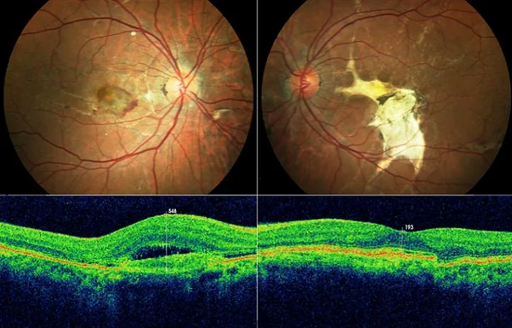
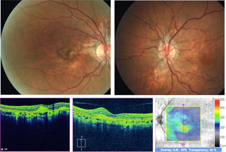
- Symptomatic presentation is usually driven by complications: metamorphopsia, central scotoma, or decreased vision from choroidal neovascularisation (CNV) involving the fovea, or sudden vision loss from choroidal rupture after even mild blunt trauma.
- May be discovered in a patient with a known systemic diagnosis (pseudoxanthoma elasticum [PXE], Paget disease, sickle cell disease) prompting screening.
- PXE patients may report characteristic skin changes — yellowish "plucked-chicken-skin" papules/plaques on the neck or flexural areas.
Findings
★ Signature Finding
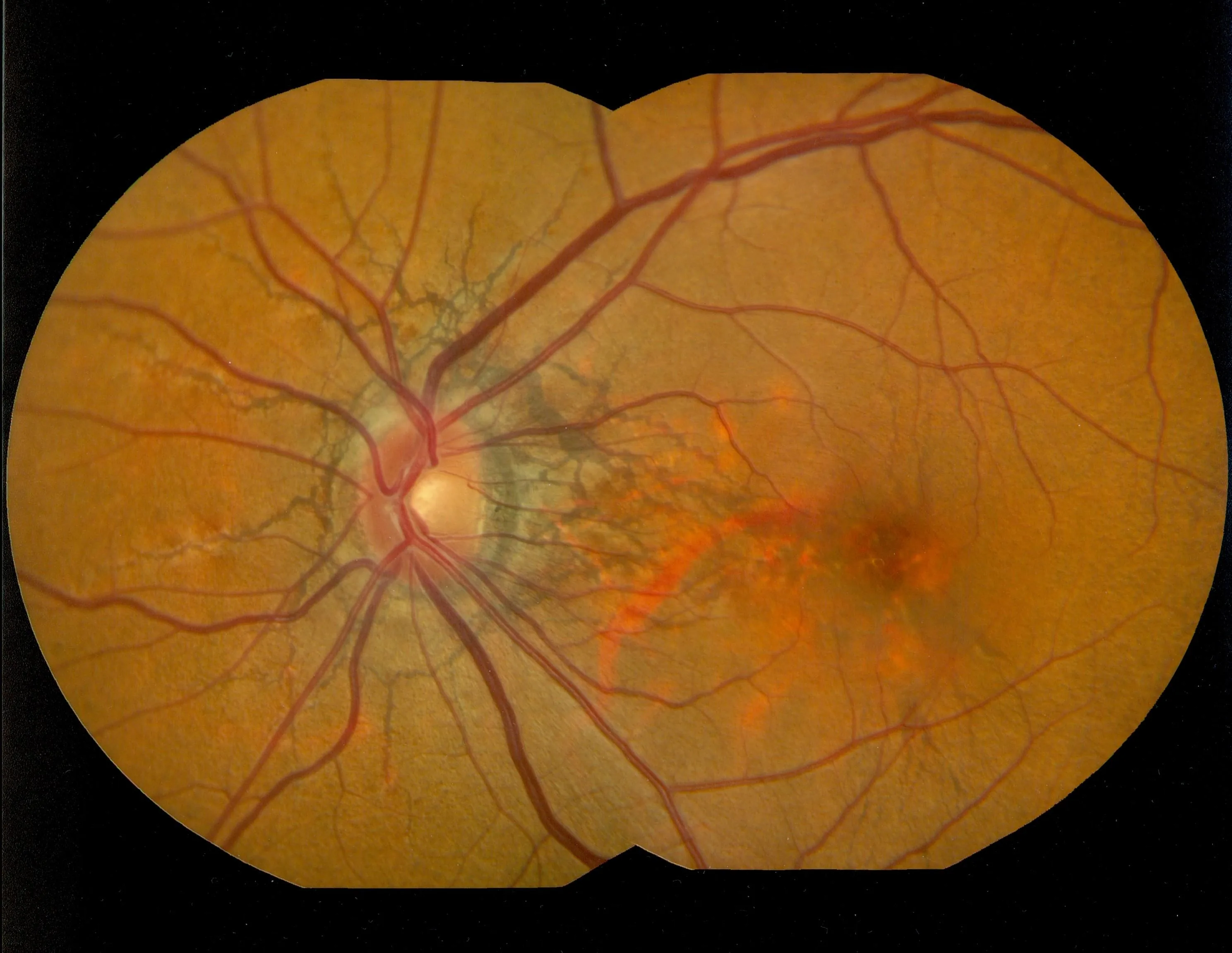
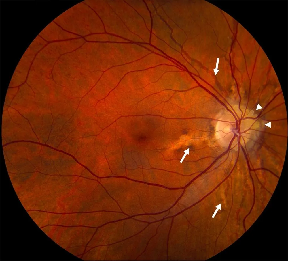
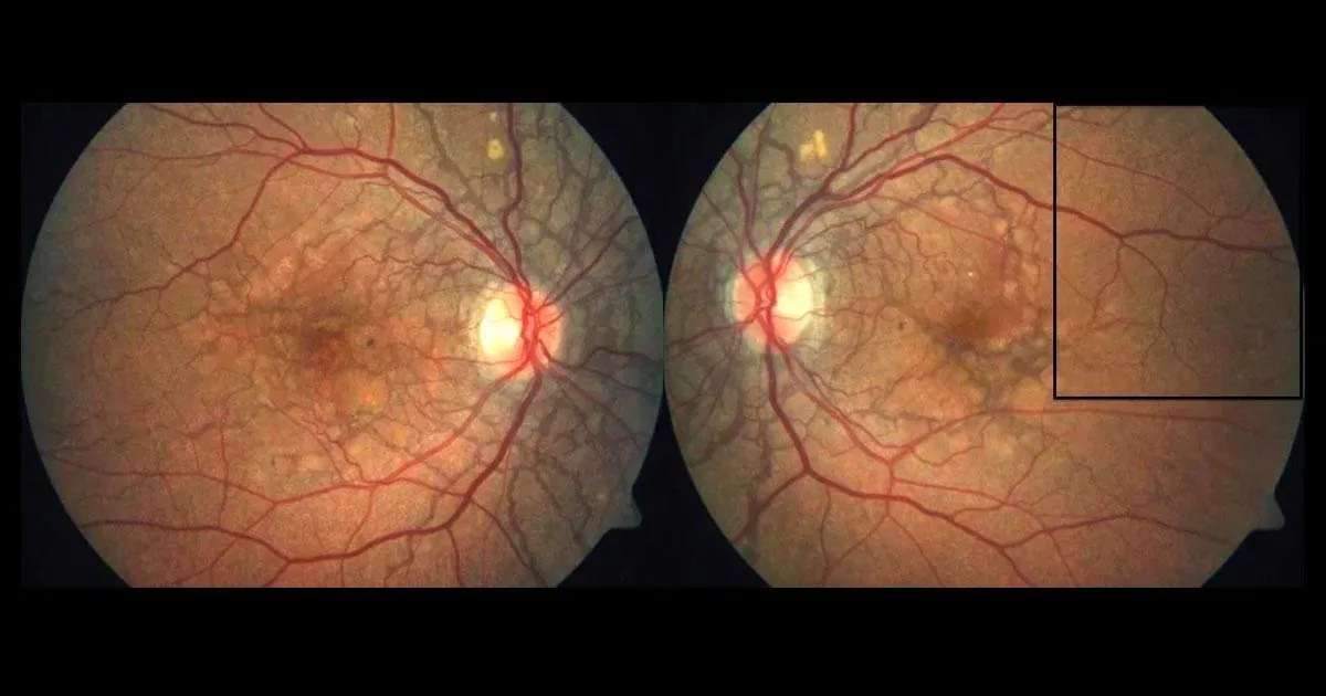
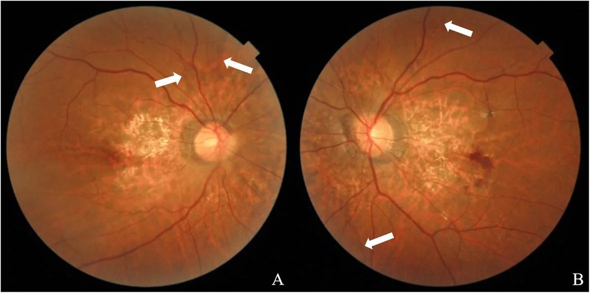
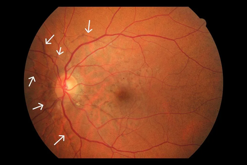
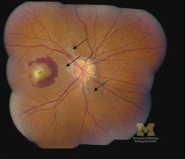
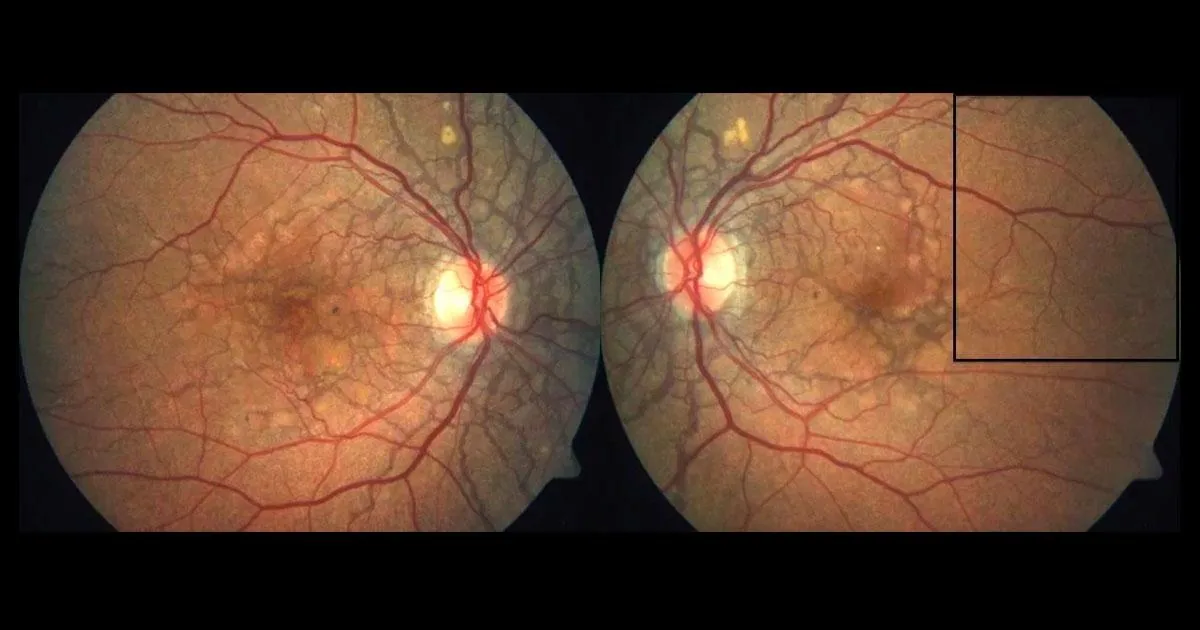
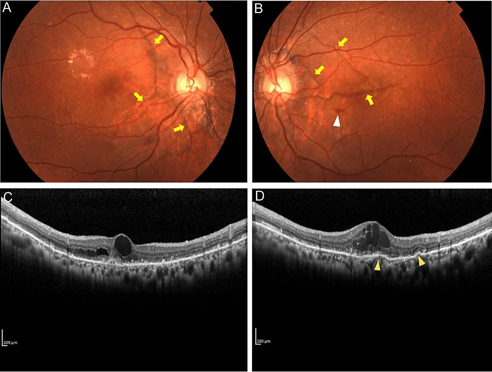
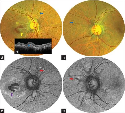
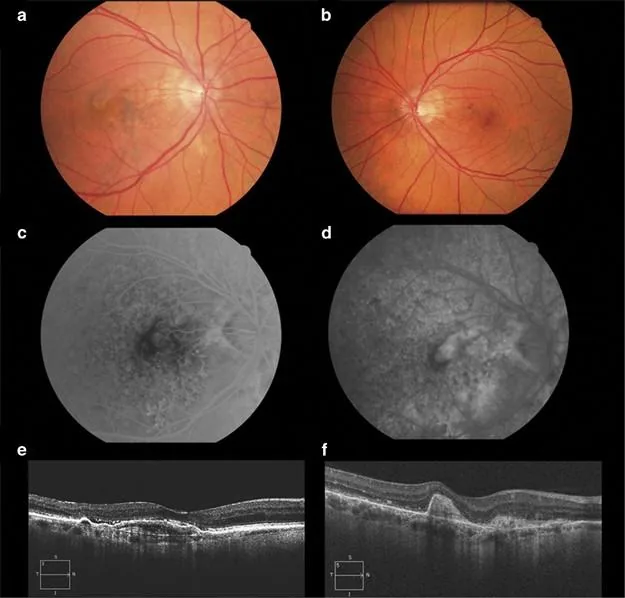
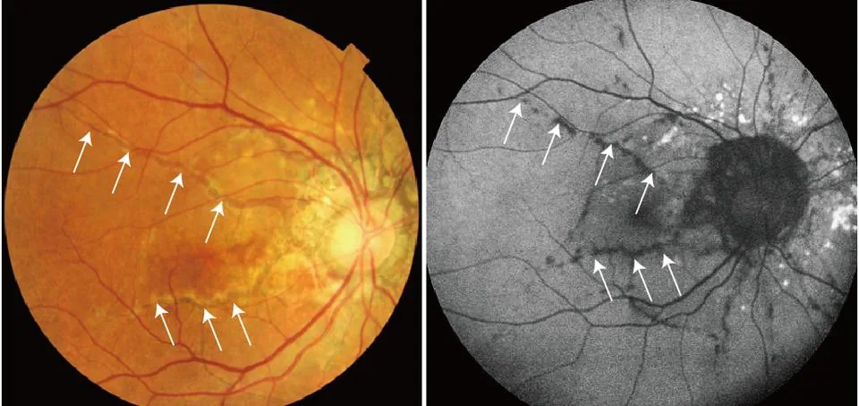
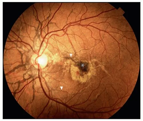
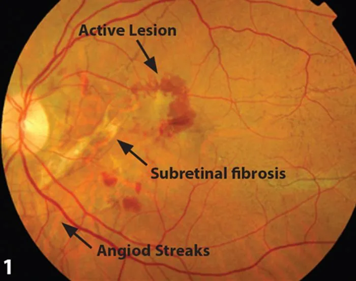
Irregular, grey-to-reddish-brown, crack-like lines radiating from the peripapillary region in a spoke- or net-like pattern, running deep to the retinal vessels. Mechanism: breaks (dehiscences) in a calcified, brittle Bruch's membrane elastic lamina, with overlying RPE atrophy.
- Peau d'orange fundus appearance (mottled peripheral pigmentary change), especially in PXE, sometimes preceding visible streaks.
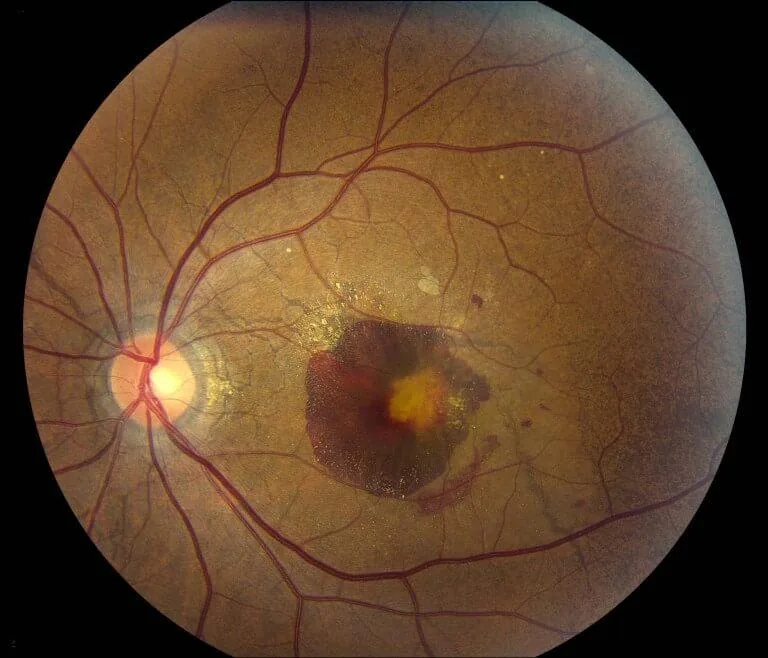
- Late complications: choroidal neovascular membrane (subretinal fluid, haemorrhage, or exudate) and subretinal/submacular haemorrhage after minor trauma.
Etiology & Mechanism
- Pathogenesis: a primary abnormality of Bruch's membrane elastic fibres, combined with increased brittleness from calcium/metallic salt deposition, leads to mechanical cracks in the membrane.
- The historical mnemonic PEPSI (Pseudoxanthoma elasticum, Ehlers-Danlos, Paget's, Sickle cell, Idiopathic) — however, contemporary evidence shows the Ehlers-Danlos association is weak (found in <1% of EDS patients in some series). The three most robust and clinically important associations are: PXE, Paget's disease of bone, and Sickle cell disease.
- Other reported associations: acromegaly, abetalipoproteinaemia, haemochromatosis, hereditary spherocytosis, and diabetes mellitus.
Red Flags — Do Not Miss
Critical — Do Not Miss
- New metamorphopsia or central vision change in a patient with known angioid streaks — urgent evaluation for CNV (the most vision-threatening complication), with prompt OCT imaging and, if confirmed, anti-VEGF therapy.
- Even minor blunt ocular trauma in a patient with angioid streaks — risk of choroidal rupture and submacular haemorrhage from the fragile Bruch's membrane; counsel against contact sports.
- New angioid streaks in a patient without a known systemic diagnosis — warrants referral for systemic workup, as the ocular finding may be the presenting sign of PXE, Paget disease, or a haemoglobinopathy.
Investigations
- Dilated fundus exam with fundus photography to document the streak pattern and monitor for change over time.
- Macular OCT (and OCT-angiography where available) to detect subclinical CNV or subretinal fluid.
- Fundus autofluorescence and fluorescein angiography if CNV is suspected.
- Systemic workup: dermatology referral/skin biopsy (PXE); serum alkaline phosphatase, calcium, phosphate (Paget disease); haemoglobin electrophoresis (sickle cell disease/haemoglobinopathies).
Management
- Routine periodic dilated fundus surveillance (with or without home Amsler grid monitoring) for early CNV detection.
- Counsel patients — especially those with PXE — to avoid activities with significant risk of blunt ocular/head trauma.
- Coordinate with internal medicine, dermatology, and/or haematology for management of the underlying systemic disease.
Treatment
- Uncomplicated angioid streaks: observation only.
- CNV secondary to angioid streaks: intravitreal anti-VEGF therapy (e.g., bevacizumab, ranibizumab, aflibercept) is the mainstay and has shown the most encouraging results of available treatments.
- Older modalities — laser photocoagulation, photodynamic therapy alone, and macular translocation — have not produced significant sustained visual improvement and are largely of historical interest for this indication.
Follow-up
- Regular dilated exam/OCT surveillance (commonly every 6–12 months, more frequently with a history of CNV) for life, given the chronic risk of neovascularisation.
- More frequent follow-up (weeks) during active anti-VEGF treatment, following standard neovascular-disease protocols.
- Ongoing coordination of systemic disease monitoring for associated conditions (e.g., cardiovascular monitoring in PXE given vascular calcification risk).
Clinical Pearl
💡 Clinical Pearl
Although the classic PEPSI mnemonic includes Ehlers-Danlos syndrome, contemporary evidence shows this association is weak; pseudoxanthoma elasticum, Paget's disease of bone, and sickle cell disease remain the three systemic associations that should actually drive the diagnostic workup.
Differential Diagnoses
| Condition | Key Distinguishing Point |
|---|---|
| Lacquer cracks of pathologic myopia | Similar linear streaks but occur with significant myopia and posterior staphyloma, not Bruch's membrane calcification. |
| Traumatic choroidal rupture | Typically a single, concentric crescentic streak around the disc after significant blunt trauma, not multiple radiating streaks. |
| Age-related macular degeneration | Yellow deposits/RPE atrophy pattern distinct from linear cracks, in an older population without the radiating pattern. |
| Choroidal folds | Parallel horizontal striae associated with an orbital mass, hypotony, or papilloedema — not a radiating crack pattern. |
| Best vitelliform macular dystrophy | Central "egg-yolk" lesion, not linear streaks; different age of onset and inheritance pattern. |