Presentation
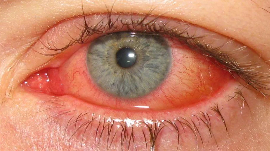
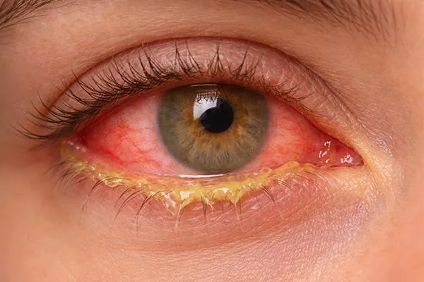
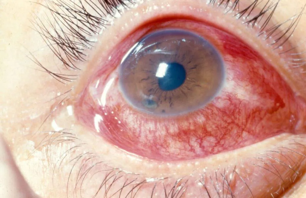
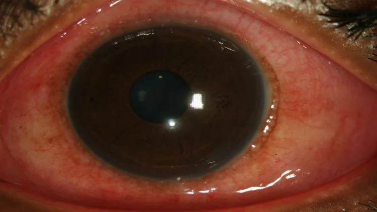
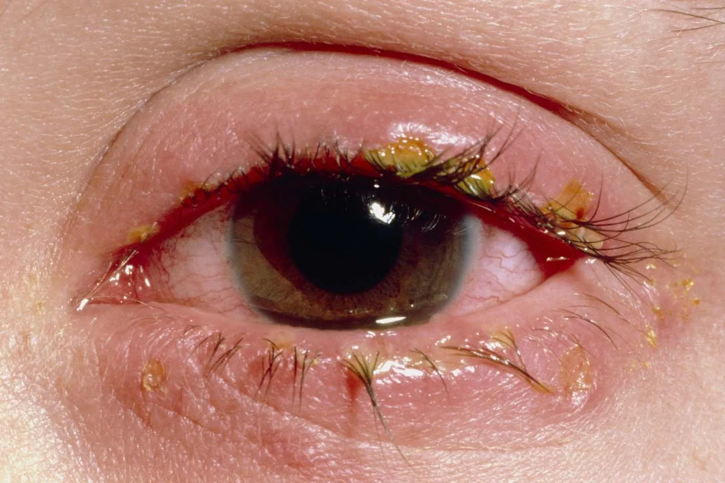
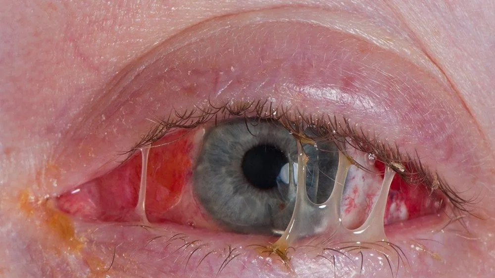
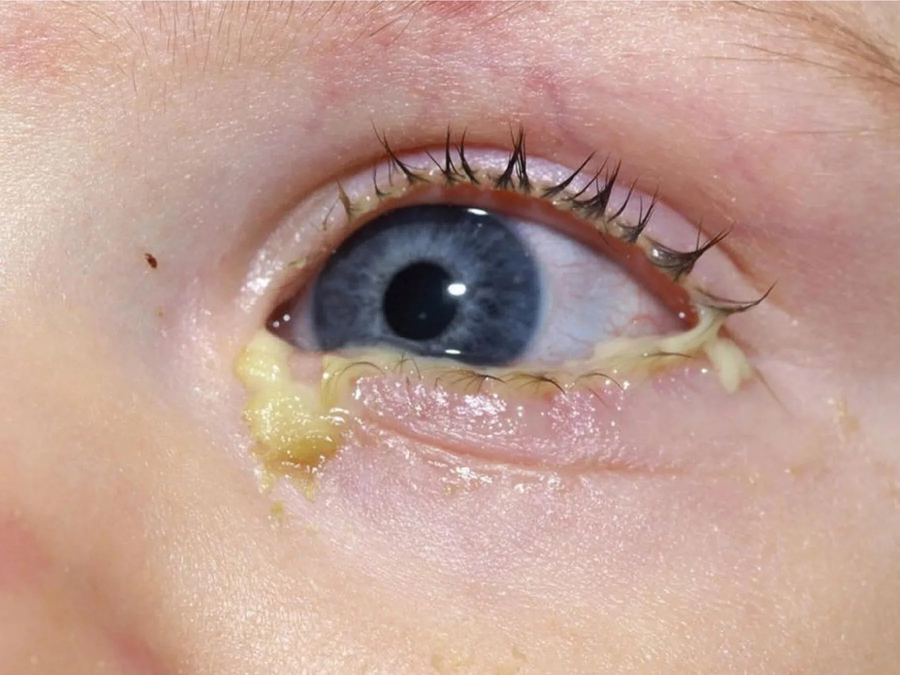
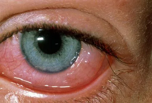
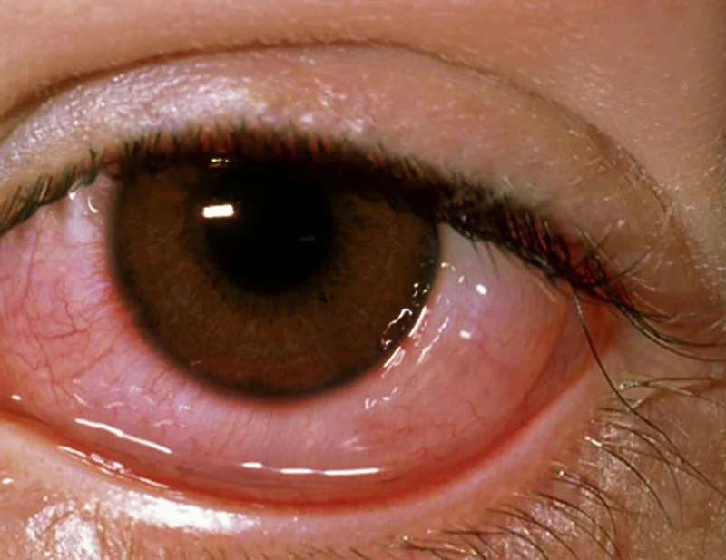
- Red eye(s) with discharge — watery discharge suggests viral or allergic aetiology; purulent/mucopurulent discharge suggests bacterial.
- Foreign-body sensation; "gluing" of the eyelids on waking is classically described with bacterial conjunctivitis.
- Viral: often bilateral (may start unilateral and spread), history of upper respiratory infection or sick contact, watery discharge.
- Allergic: bilateral itching (hallmark symptom), watery discharge, seasonal pattern or personal/family history of atopy.
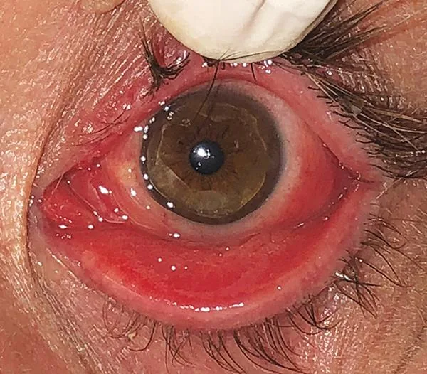
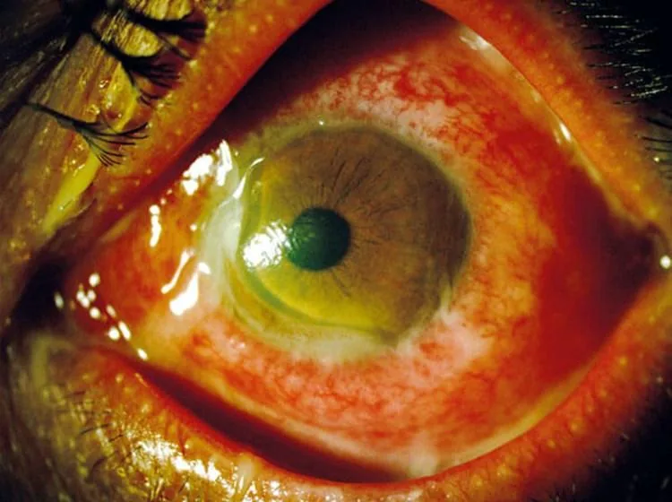
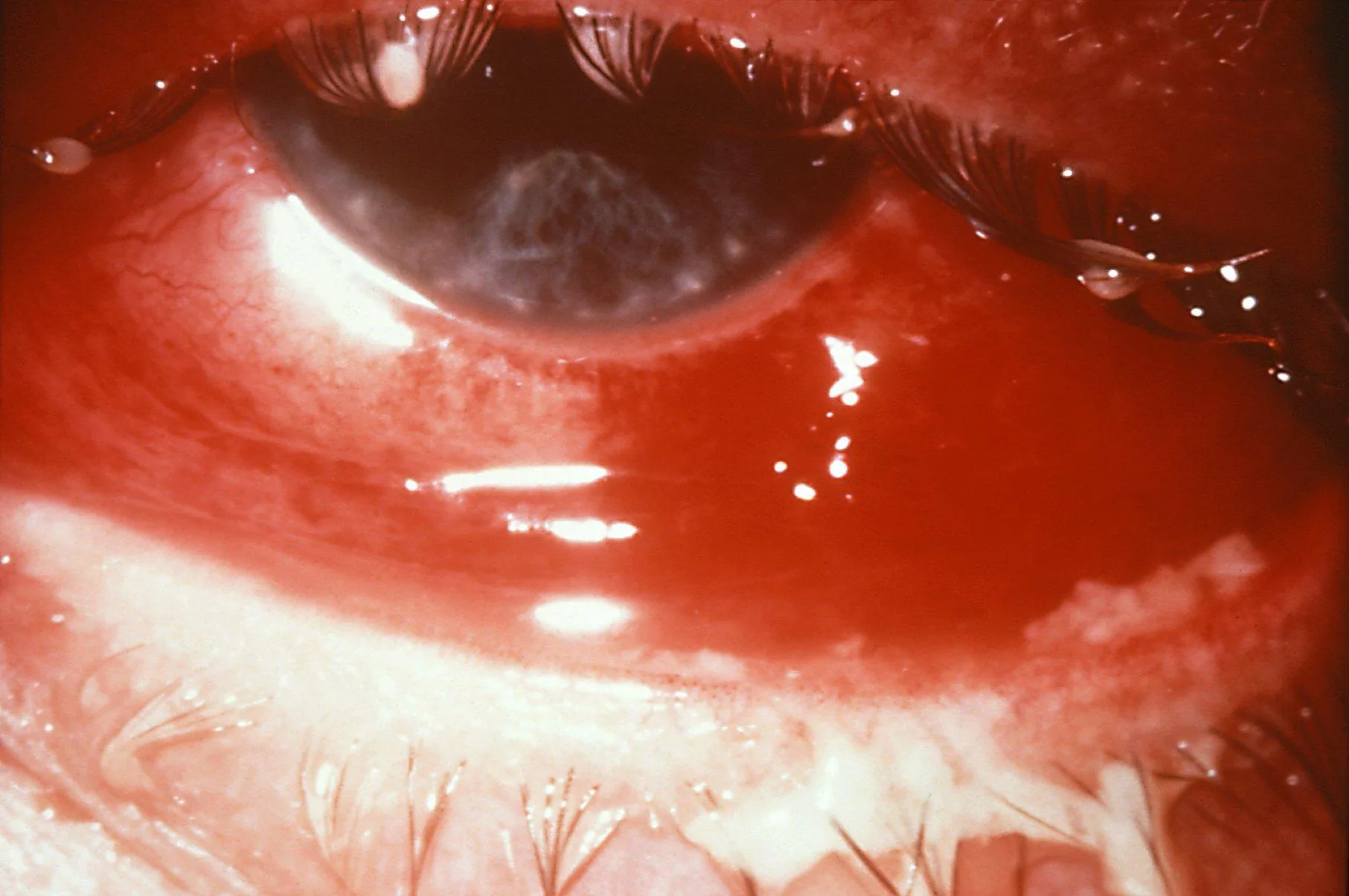
- Hyperacute (gonococcal): rapid-onset copious purulent discharge and marked chemosis, history of sexual exposure or maternal infection in a neonate — a true ocular emergency due to corneal perforation risk.
Findings
★ Signature Finding
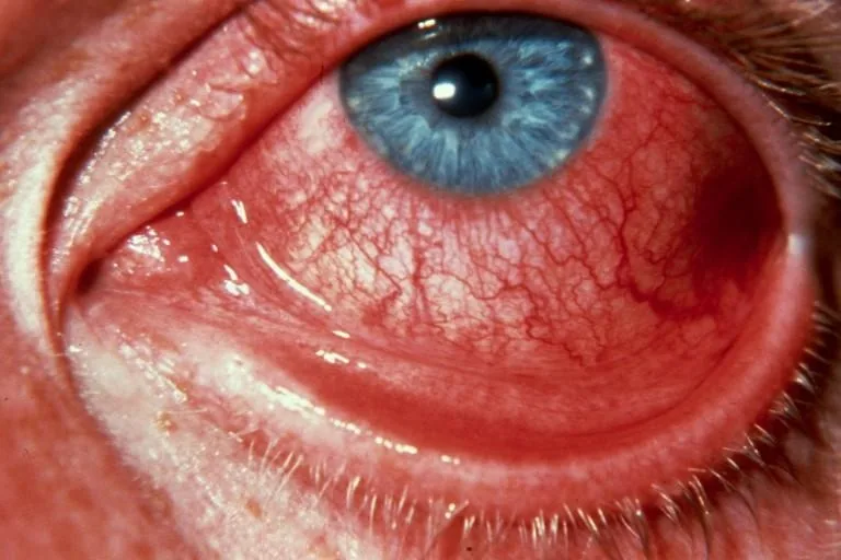
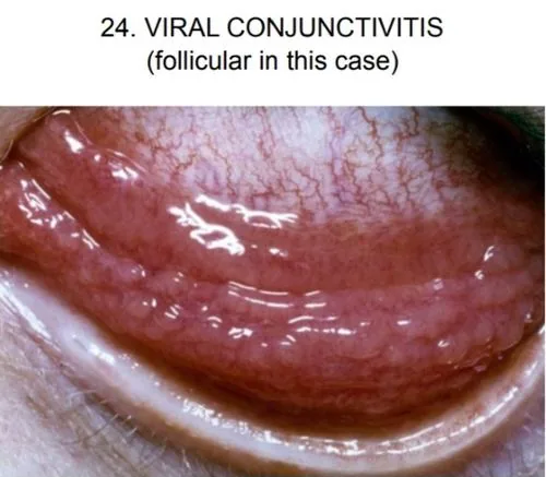
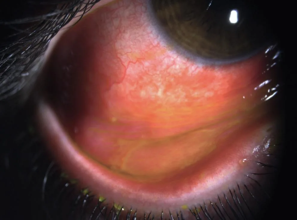
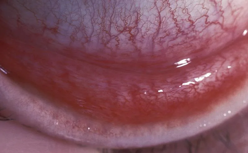
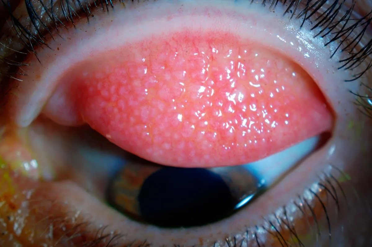
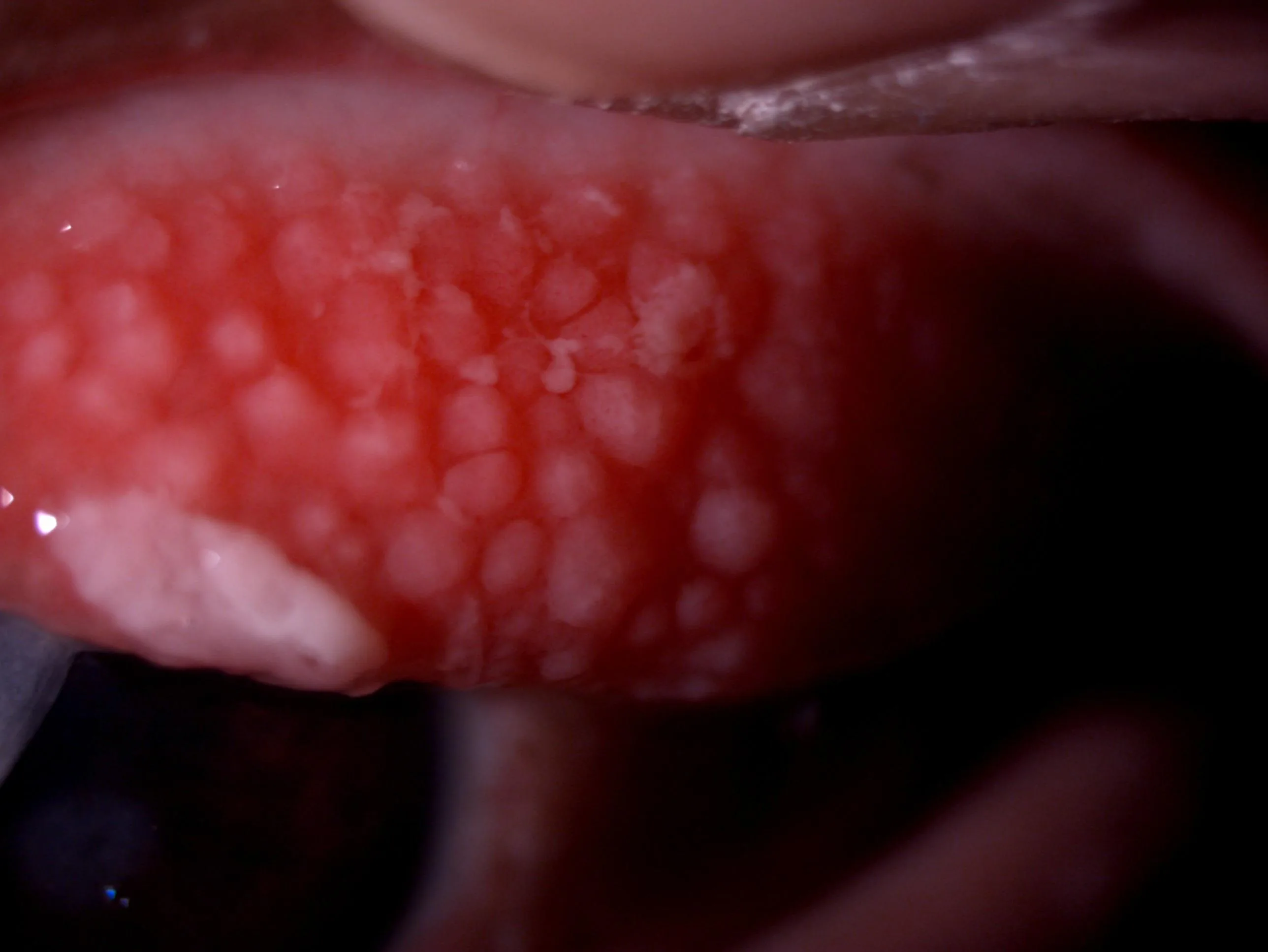
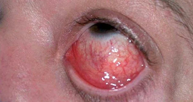
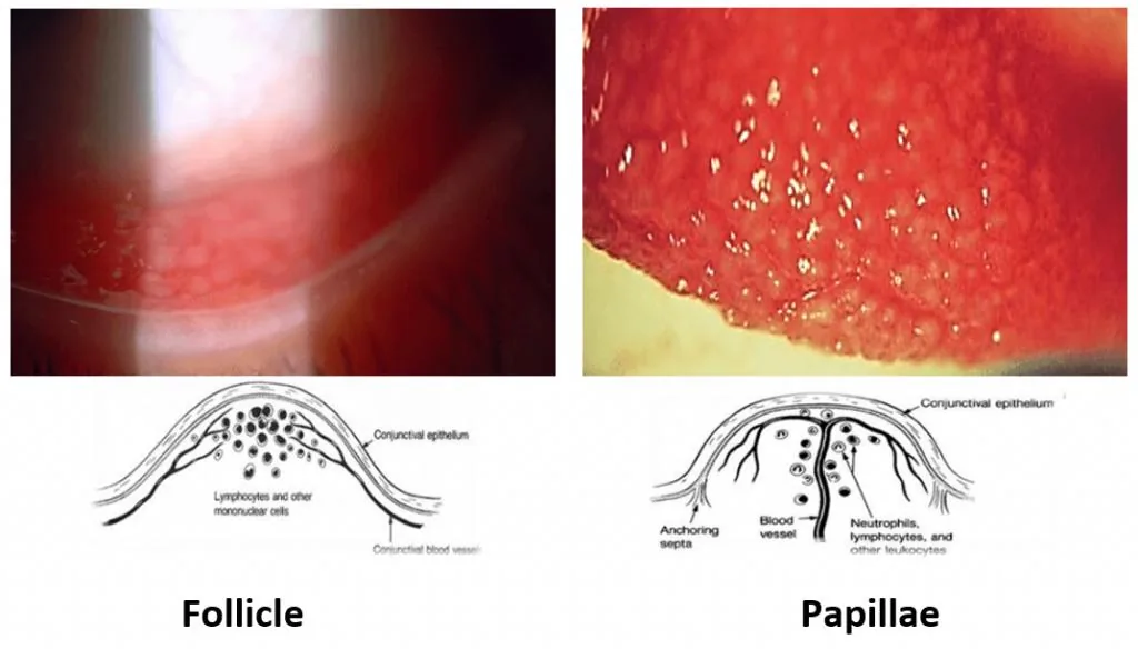
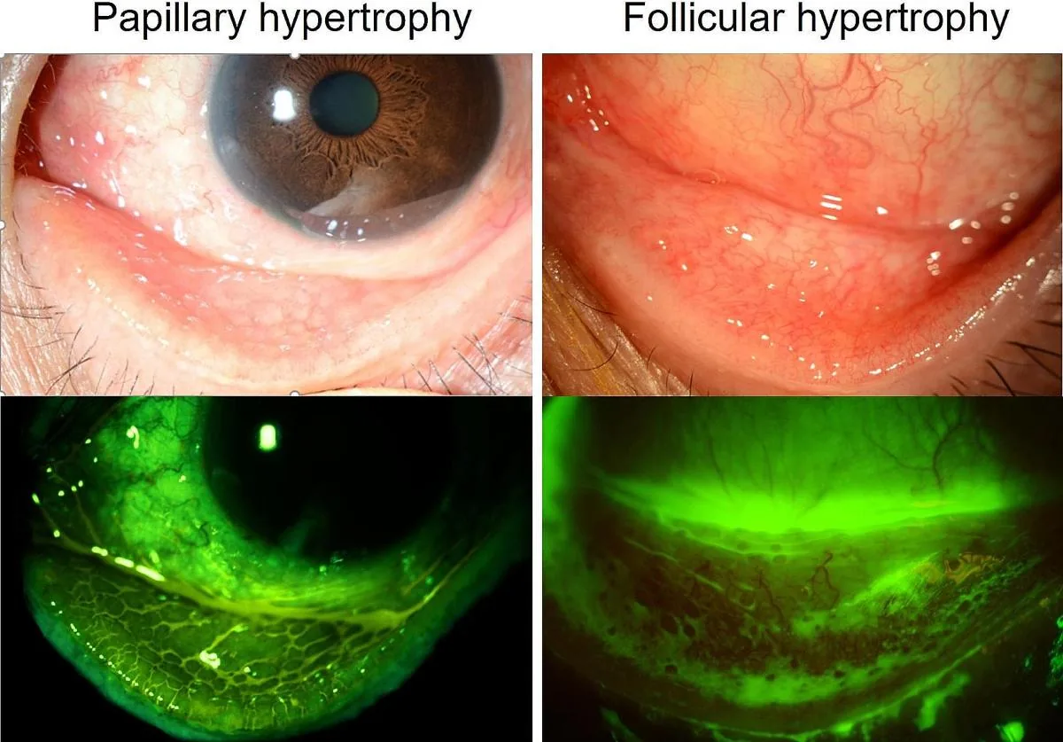
Viral (adenoviral): follicular conjunctival reaction with preauricular lymphadenopathy (reported in up to half of viral cases). Mechanism: reactive lymphoid hyperplasia of conjunctiva-associated lymphoid tissue.
- Epidemic keratoconjunctivitis (severe adenoviral subtype): watery discharge, hyperaemia, chemosis, ipsilateral preauricular lymphadenopathy, later subepithelial infiltrates.
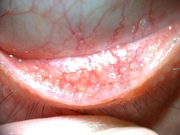
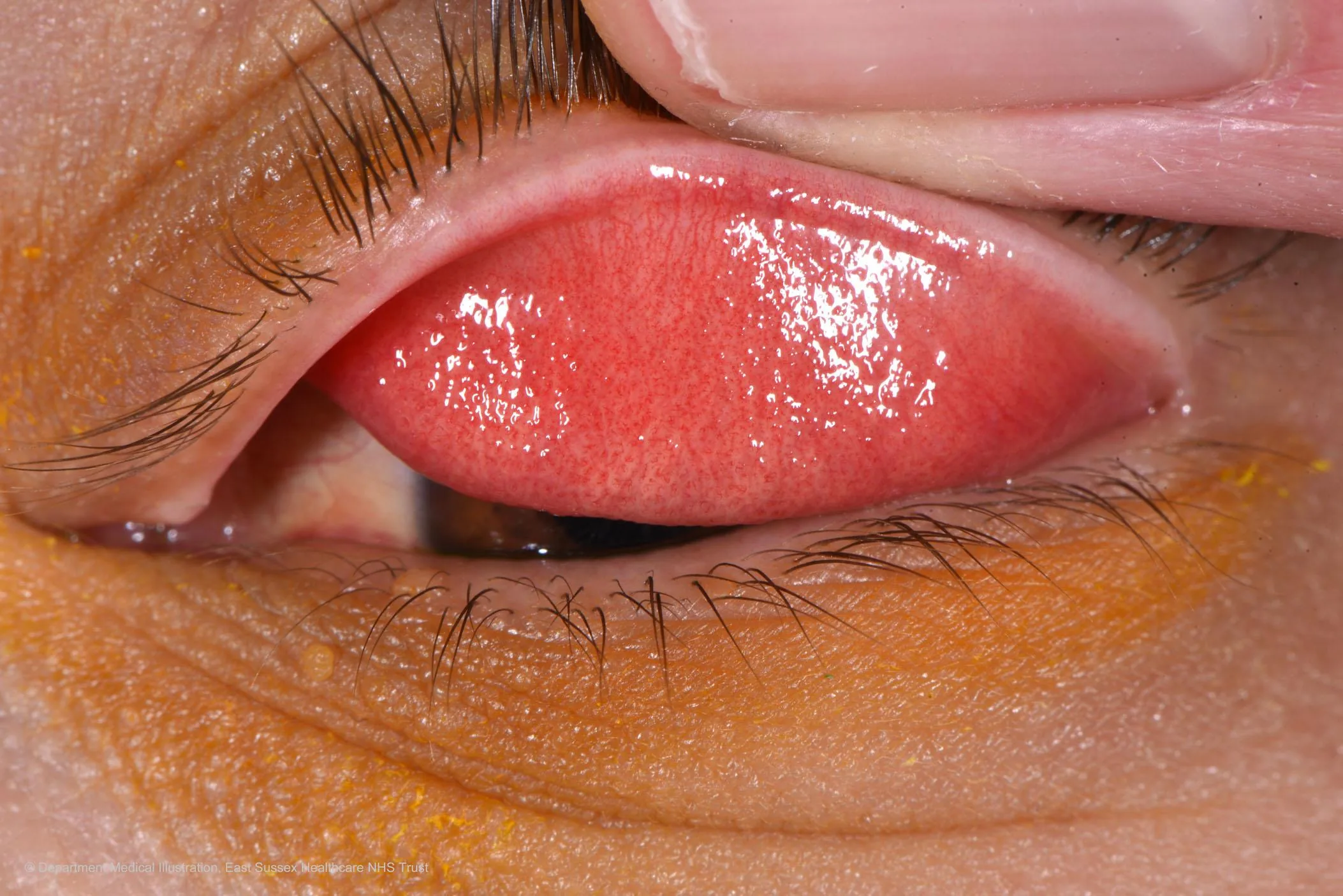
- Bacterial: papillary conjunctival reaction, mucopurulent discharge, usually without significant lymphadenopathy (except hyperacute gonococcal/chlamydial disease).
- Allergic: papillary reaction with chemosis; often concurrent allergic rhinitis.
Etiology & Mechanism
- Viral is the most common cause overall; adenovirus predominates. Certain serotypes (3, 4, 7) are linked to pharyngoconjunctival fever; others (8, 19, 37, 53, 54) to epidemic keratoconjunctivitis.
- Bacterial: Staphylococcus aureus, Streptococcus pneumoniae, and Haemophilus influenzae most common; hyperacute disease classically caused by Neisseria gonorrhoeae; chronic/hyperacute also by Chlamydia trachomatis.
- Allergic: IgE-mediated mast cell degranulation in seasonal/perennial disease; T-cell/eosinophil-mediated in vernal and atopic keratoconjunctivitis.
Red Flags — Do Not Miss
Critical — Do Not Miss
- Marked purulent discharge with rapid progression, especially with risk factors for gonococcal infection — corneal involvement and perforation risk demand same-day emergent treatment.
- A contact lens wearer with conjunctivitis and pain out of proportion to exam findings — reconsider microbial keratitis or Acanthamoeba rather than simple conjunctivitis.
- Reduced vision, significant pain, photophobia, or visible corneal involvement — these are NOT features of uncomplicated conjunctivitis; evaluate for keratitis, uveitis, or angle closure.
- Neonatal conjunctivitis (ophthalmia neonatorum) — always an emergency; evaluate for gonococcal and chlamydial aetiology.
Investigations
- Diagnosis is usually clinical; further testing is not required for typical, mild presentations.
- Rapid antigen detection (e.g., adenovirus point-of-care test) can help confirm adenoviral aetiology in ambiguous cases.
- Conjunctival culture, Gram stain, and PCR reserved for hyperacute presentations, neonatal conjunctivitis, recurrent/refractory cases, or immunocompromised patients.
Management
- Viral disease: supportive care — cool compresses, artificial tears, and strict hand hygiene given high contagiousness (~10–50% for adenoviral disease).
- Discontinue contact lens wear until resolved.
- Avoid unnecessary or prolonged topical antibiotic use in confirmed/likely viral conjunctivitis.
Treatment
- Bacterial: most cases are self-limited and clear within ~10 days; topical antibiotics (e.g., a fluoroquinolone or polymyxin/trimethoprim) shorten symptom duration and speed clearance, and are generally recommended — especially for contact lens wearers.
- Hyperacute gonococcal: same-day emergent treatment — systemic (IM/IV) ceftriaxone plus saline irrigation and topical antibiotic; treat empirically for concurrent chlamydial infection.
- Allergic: topical dual-acting antihistamine/mast-cell-stabiliser agents (e.g., olopatadine), cold compresses, allergen avoidance; oral antihistamines as adjunct.
- Viral (adenoviral): primarily supportive; topical corticosteroids reserved for visually significant subepithelial infiltrates or membrane formation, only after HSV keratitis has been excluded.
Follow-up
- Bacterial: recheck if not improving within a few days of starting antibiotics.
- Viral: monitor for subepithelial infiltrates, which can appear 1–3 weeks after onset in adenoviral EKC and may require a slowly tapered topical steroid course.
- Reinforce hygiene and contagion-period counselling at every visit.
Clinical Pearl
💡 Clinical Pearl
Preauricular lymphadenopathy is present in up to half of viral conjunctivitis cases and is distinctly uncommon in typical acute bacterial conjunctivitis (aside from hyperacute gonococcal disease) — a useful bedside discriminator alongside discharge character.
Differential Diagnoses
| Condition | Key Distinguishing Point |
|---|---|
| Acute angle-closure glaucoma | Pain, elevated IOP, mid-dilated pupil, corneal oedema — all absent in simple conjunctivitis. |
| Anterior uveitis | Photophobia, ciliary flush, cells/flare in the anterior chamber, minimal discharge. |
| Episcleritis/scleritis | Localised/sectoral injection; scleritis is markedly tender to palpation; no discharge. |
| Dry eye disease | Chronic, often bilateral, minimal discharge, abnormal tear film testing. |
| Microbial keratitis | Corneal infiltrate, pain out of proportion, decreased vision, often contact-lens related. |