


Presentation
- Contact lens wearer presenting with pain that is often out of proportion to early clinical findings, photophobia, redness, foreign-body sensation, and blurred vision.
- History often includes water exposure while wearing lenses — swimming, showering, or rinsing lenses/case with tap water.
- Frequently misdiagnosed early as herpes simplex keratitis because early epithelial changes can look dendritiform.
Findings
★ Signature Finding
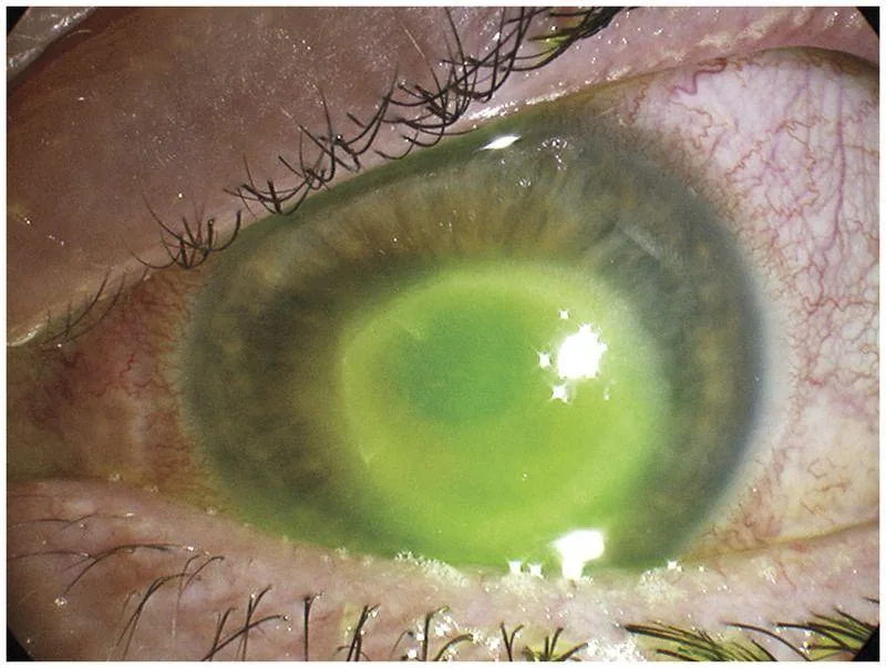
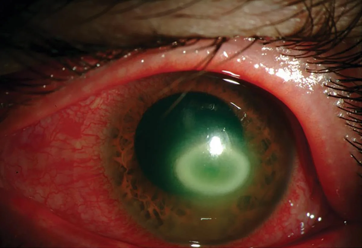
Stromal ring-shaped infiltrate (a late finding). Mechanism: immune-mediated inflammatory response to amoebic antigens, often centred on the perineural/stromal tissue.
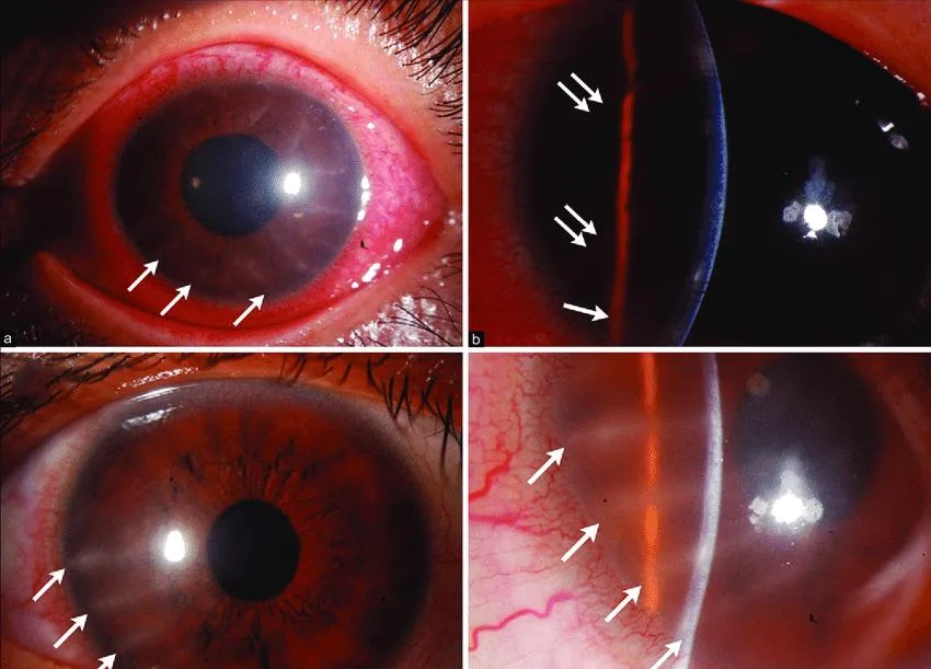
- Radial perineural (perineuritis) infiltrates — inflammation tracking along corneal nerves — an earlier, relatively specific sign.
- Early epithelial pseudodendrites can mimic HSV keratitis.
- Late disease: dense ring infiltrate, stromal haze/scarring; can progress to scleritis in severe or delayed cases.
Etiology & Mechanism
- Acanthamoeba is a ubiquitous free-living protozoan found in soil and water, existing in an active trophozoite form and a dormant, biocide-resistant cyst form.
- Pathogenesis requires corneal trauma/epithelial defect plus exposure to the organism (commonly via contaminated water or lens solution).
- Risk factors: contact lens wear with poor hygiene or water exposure; in non-lens wearers, corneal trauma followed by exposure to contaminated water/soil.
Red Flags — Do Not Miss
Critical — Do Not Miss
- Pain disproportionate to signs in a contact lens wearer with water exposure history — do not anchor on a bacterial or herpetic diagnosis.
- Radial perineural infiltrates or a ring infiltrate — treat empirically for Acanthamoeba rather than waiting for confirmatory culture, since delayed anti-amoebic therapy worsens outcomes.
- Poor response to standard antibacterial or antiviral therapy in a red, painful eye with a contact lens history.
Investigations
- In-vivo confocal microscopy to visualise cysts — rapid, non-invasive.
- Corneal scraping with KOH + calcofluor white staining to visualise double-walled cysts.
- Culture on non-nutrient agar overlaid with E. coli (Page's/Neff's agar).
- PCR of corneal scrapings, increasingly used for confirmation.
- Corneal biopsy if scraping is non-diagnostic but clinical suspicion remains high.
Management
- Start empirical anti-amoebic therapy on clinical suspicion — do not wait for culture confirmation given the time-sensitivity of the disease.
- Epithelial debridement to reduce organism load and improve drug penetration.
- Discontinue contact lens wear.
Treatment
- First line: a cationic antiseptic biguanide — PHMB 0.02% or chlorhexidine 0.02% — often combined with a diamidine (propamidine 0.1% or hexamidine), dosed intensively (e.g., hourly) initially, then tapered over weeks to months.
- No medication is formally approved for this indication in most countries; treatment is off-label.
- Topical corticosteroids are controversial: may reduce stromal inflammatory scarring later in the course but risk worsening amoebic proliferation if started too early.
- Refractory disease: therapeutic penetrating keratoplasty deferred until medical cure achieved; adjunctive photodynamic therapy with Rose Bengal has shown encouraging results in a small pilot series.
Follow-up
- Frequent early follow-up (days) to judge treatment response.
- Total treatment courses are often prolonged — months, not weeks — given cyst resistance; taper slowly to reduce recurrence risk.
- Monitor for scleritis and secondary glaucoma throughout treatment.
Clinical Pearl
💡 Clinical Pearl
Because early Acanthamoeba keratitis can look like HSV keratitis (pseudodendrites), persistent pain and lack of response to antivirals in a contact lens wearer should trigger a dedicated anti-amoebic workup rather than a longer antiviral trial.
Differential Diagnoses
| Condition | Key Distinguishing Point |
|---|---|
| HSV keratitis | True dendrites with terminal bulbs and reduced corneal sensation; Acanthamoeba often has preserved/increased sensation early with radial perineural infiltrates. |
| Bacterial keratitis | More rapid onset, purulent discharge, responds to standard antibiotics. |
| Fungal keratitis | Feathery infiltrate margins, satellite lesions, history of vegetable-matter trauma. |
| Microsporidial keratoconjunctivitis | Occurs in immunocompromised hosts, diffuse punctate epitheliopathy. |
| Herpes zoster keratitis | Dermatomal vesicular rash, history of shingles. |
References
- EyeWiki. Acanthamoeba Keratitis.
- Herz NL, et al. Comparison of PHMB and Chlorhexidine as Monotherapy Agents in the Treatment of Acanthamoeba Keratitis. Am J Ophthalmol.
- Chlorhexidine 0.02% or PHMB 0.02% as the main treatment for Acanthamoeba keratitis. PubMed.
- Outcome of photodynamic therapy with Rose Bengal in conjunction with topical PHMB and chlorhexidine in Acanthamoeba keratitis. PMC.