Active Cross-Section Meridian:
Refraction Simulator & Physiological Optics
Educational simulator — not a medical device.
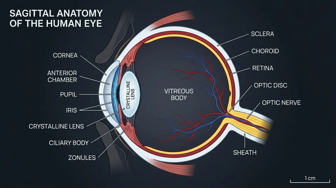
In an emmetropic eye, parallel light rays from infinity are refracted by the cornea (~43 D) and crystalline lens (~17-20 D) so that they converge exactly onto the fovea centralis of the retina (axial length ~24.0 mm) without requiring any accommodative effort.
Light rays converge to a focal point in front of the retina. This occurs either because the eyeball is too long (axial myopia) or the optical elements refract too strongly (refractive/curvature myopia). Diverging past the focal point, rays hit the retina as a blurred blur circle. Corrected by a concave (divergent / minus) lens that moves the focal point back onto the retina.
Light rays converge toward a virtual point behind the retina because the eye is either too short (axial hyperopia) or too weak (refractive hyperopia). A convex (convergent / plus) lens adds positive vergence to bring the focal point forward onto the retina.
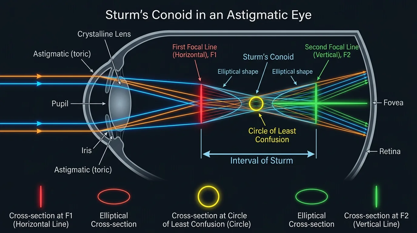
The cornea or lens possesses different refractive powers along two perpendicular meridians, creating an aspheric interval of Sturm between two distinct focal lines. Spherocylindrical lenses (combining spherical and cylindrical curves at a specific axis) neutralize both meridians simultaneously.